Super Resolution Microscopy of Nuclear Chromatin: What Advantages will Quantum Enhanced Microscopy Bring?

TIME: 1:00pm
WHEN: 4 June, 2025
LOCATION: Zoom
TIMEZONE: AEST
Join us for this intriguing QUBIC Seminar Series event.
Super Resolution Microscopy of Nuclear Chromatin: What Advantages will Quantum Enhanced Microscopy Bring?
Speaker: Dr Mike Mlodzianoski, UoM
Date: Wednesday 04 June, 1pm – 2pm AEST
Zoom: Click here to join the seminar
Abstract: DNA within the nucleus is organized into nucleosomes, which are clustered into chromatin. These structures are continuously undergoing rearrangement to enable proteins access to different sections of DNA for reading and copying genetic information. The nucleosome and chromatin cluster on spatial scales of tens and hundreds of nanometres, respectively, much smaller than the diffraction limit of light, making them ideal candidates to be studied with single molecule localization microscopy. I will present recent results of our super resolution microscopy experiments studying chromatin in live and fixed cells and the difficulties of imaging in the nucleus. I will discuss how quantum enhanced super resolution approaches and Hong-Ou-Mandel fluorescence lifetime imaging microscopy can reveal new information about chromatin structure and dynamics.
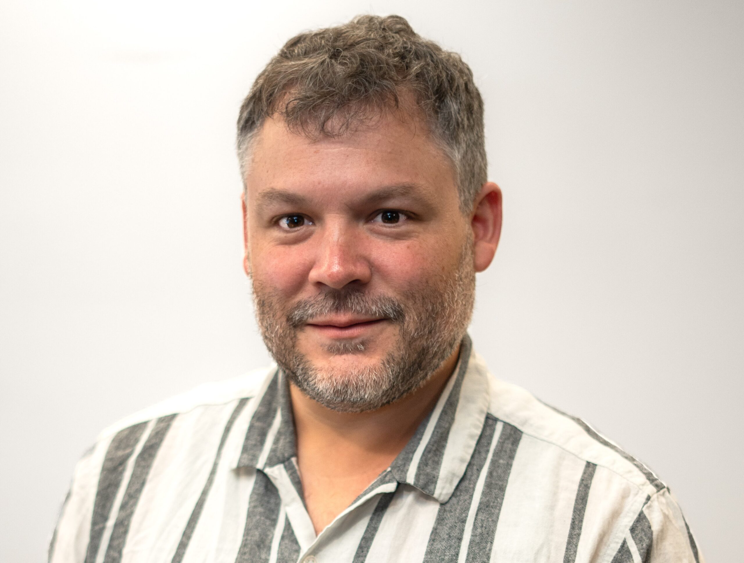
Bio: Dr Michael Mlodzianoski is a Postdoctoral Fellow in the lab of A/Prof Elizabeth Hinde in the School of Physics at the University of Melbourne and a member of QUBIC. Throughout his career Michael has made many significant contributions to the development of new techniques in single molecule localization microscopy. During his PhD at the University of Maine, Michael developed a single molecule localization method that measures both the super-resolved spatial position and fluorescence spectra of single molecules. During his first Postdoctoral position, Michael pioneered a cutting-edge implementation of adaptive optics in a single molecule localization system to remove optical aberrations to enable highly accurate and precise 3D super resolution measurements in thick tissue samples. Michael is currently working on applying super resolution microscopy to the study of chromatin structure and dynamics while also developing quantum enhanced super resolution and fluorescence lifetime imaging microscopes.
Email CoordinatorView all events