Unlocking the Hidden Brain with Quantum Technology: A New Era of Brain Measurement

When Curiosity Meets Brain Science

Dr Sophie Lin
Dr Sophie Lin

When Sophie Lin talks about the brain, her tone is equal parts wonder and humility. “The more you study the brain, the less you feel like you really understand it,” she says – a line that neatly captures both her scientific motivation and her career path. Trained originally as a neurologist, she encountered a gap between what brain images indicated and what patients with epilepsy, stroke, and degenerative diseases experienced. The brain imaging story didn’t quite add up. So she crossed the bridge into academia, determined to ask deeper questions about how the brain actually works.

Today, Sophie manages the optically pumped magnetoencephalography (OP-MEG) facility at the University of Melbourne – Australia’s first whole-head, wearable imaging system of its kind – and is part of a broader effort to use quantum sensing technologies to image biological systems in new and more precise ways. Her focus is a part of the brain that has long been overlooked: the cerebellum.

The Hidden Brain
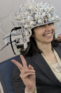
Sophie’s colleague QUBIC CI Prof Marta Garrido

The cerebellum sits at the bottom of the brain – tucked away, hard to reach, and even harder to measure. The magnetic signals the brain emits are billions of times weaker than the Earth’s magnetic field – it’s like trying to hear a whisper in the middle of a stadium concert. For a long time, the cerebellum was thought of as a supporting actor, mainly associated with movement and coordination. Increasingly, patient data and neuroscience studies suggest it plays a much more important role, including in language, learning, and higher-order cognition. But to understand what it really does, researchers first need a reliable way to observe it in action.

For most of modern neuroscience, that has been the hard part. Traditional MEG systems rely on sensors that must be cooled to extremely low temperatures and housed in large, rigid, shielded rooms. The hardware sits several centimetres away from the scalp, which limits sensitivity – especially for deeper brain structures like the cerebellum.

The result has been a neuroscience blind spot. In clinical practice, Sophie saw the consequences of that gap firsthand: patients whose symptoms could not be cleanly explained by what standard imaging showed. As she puts it, structure alone does not always tell you how the brain is functioning in real time.

For years, the implicit conclusion was simple: measuring cerebellar activity non-invasively is too difficult to do well. That assumption is now being challenged by a new class of quantum sensors – and that could lead to a better understanding of neurodegenerative diseases, stroke recovery, and epilepsy.

Enter Quantum Sensors
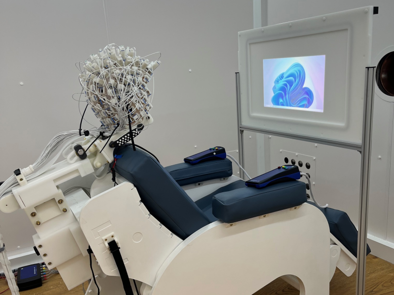
The MBCIU OPM-MEG system provides researchers access to an unparalleled suite of imaging technology for structural and functional brain research

OP-MEG uses optically pumped magnetometers – quantum sensors that operate at room temperature. Instead of being locked into a fixed helmet inside a bulky cryogenic system the size of a refrigerator, these sensors can be placed directly on the head, in a lightweight, wearable array. For researchers like Sophie, that significantly improves what is possible in neuroscience imaging.

This proximity matters. By bringing the sensors much closer to the scalp, OP-MEG can capture brain signals that are two to three times stronger than those measured by conventional systems. The sensors are mounted in a wearable helmet that can be customised from an individual’s MRI scan, allowing researchers to position sensors closer to regions of interest such as the cerebellum. This personalised design also makes scanning more accessible – enabling studies with children or elderly participants who may struggle to remain completely still.

In Sophie’s work, this quantum sensing platform is doing something radical: it is giving the cerebellum “another chance” to be studied properly. This is not about a single disease or a single experiment. It is about building a new measurement capability – a platform technology that lets researchers observe parts of the brain that have been, until now, largely hidden.

This is quantum technology in action: not just a laboratory curiosity, but a tool that changes what can be measured, who can be measured, and what questions can be asked – building on decades of animal research to show that wearable quantum sensors can now detect cerebellar activity in humans.

From Discovery to Helping People Speak Again

The implications of this new window on the brain are wide-ranging. Better access to cerebellar signals could deepen understanding of conditions such as stroke, alcohol-related brain damage, neurodegenerative disorders, and psychiatric disorders. It could reshape how scientists think about sleep, learning, and even consciousness. Increasingly, evidence suggests the cerebellum is involved in far more than movement – but without the right tools, those hypotheses have been difficult to test.

One of Sophie’s current directions is exploring how the cerebellum contributes to language and speech production. If researchers can identify reliable neural markers linked to how the brain produces language, those signals could one day be used to build more advanced, brain-informed speech assistance technologies for people who struggle to speak because of neurological conditions.

By turning the cerebellum from a blind spot into a field of discovery, OP-MEG is expanding what brain science can do, and showing how Australia’s quantum capability can translate into tools that change how we explore the most complex system we know: the human brain.

visit the Cognitive Neuroscience and Computational Psychiatry Laboratory

visit the Melbourne Brain Centre Imaging Unit (MBCIU)
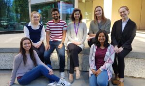
Team photo: Cognitive Neuroscience and Computational Psychiatry Laboratory
The Cognitive Neuroscience and Computational Psychiatry Laboratory team at University of Melbourne
Launch OPM-MEG facility
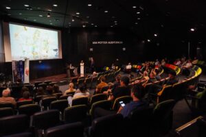
Launch of the Optically Pumped Magnetometer MEG (OPM-MEG) facility in March 2026

Seeing Dementia Unfold

Dementia: a growing challenge, with limited answers
image Lezanne Ooi
“Quantum sensors can detect neuronal signals and molecular-scale changes with extreme sensitivity, measuring subtle changes in neurons and identifying disease-specific molecular fingerprints.” — Chief Investigator Prof Lezanne Ooi

Dementia is one of Australia’s most pressing health challenges, and the second leading cause of death in Australia. Beyond the statistics lies a deeply personal toll on
individuals, families, carers and communities.

Work underway at QUBIC is opening a new window into dementia, using quantum sensing to observe brain cells in ways that were not previously possible.

Despite decades of dementia research, there is still no cure. Recently approved medications can slow symptoms for some people, but they are not suitable for everyone. Some require regular MRI scans to monitor serious side effects.

While these treatments may slow symptoms for some people, they do not stop or reverse the underlying disease, meaning brain cells continue to be irreversibly lost.

A major challenge in dementia research is understanding how the disease begins and progresses. Scientists typically compare healthy brain cells with diseased ones, looking for differences that might explain why neurons fail and die. However, this approach captures only snapshots of a disease that develops over years or decades, missing how a healthy neuron gradually becomes diseased as damage accumulates. Part of the challenge lies in the limits of existing microscope technology, which offers low-resolution photographs. What’s needed is a high-resolution movie, showing how cells change and interact over time.

Growing the human brain in a dish

QUBIC Chief Investigator and Deputy Director Professor Lezanne Ooi is working to overcome this barrier. A group leader at the University of Wollongong and Deputy Director of the Molecular Horizons Research Institute, Lezanne leads a research program grounded in cellular neuroscience – the study of how individual brain cells function, communicate and fail.

At the core of her work is a powerful platform technology. Using a small skin sample donated by a patient, Lezanne’s team reprograms those cells into stem cells, and then guides them to become human brain cells grown in a dish.

Crucially, these cells are alive, accessible and measurable, opening new possibilities for understanding disease mechanisms and testing potential therapies, without
needing to sample a patient’s brain tissue.

While dementia is a central focus, Lezanne’s work also spans Parkinson’s disease, motor neuron disease, epilepsy and other rare brain diseases, as well as emerging
questions around genetic and environmental risk factors for neurodegenerative diseases.

Where quantum sensing changes the picture

Advances in quantum sensing are opening new possibilities in biology and medicine, allowing researchers to probe living systems with unprecedented sensitivity.

Quantum sensors are exquisitely sensitive to tiny electrical and magnetic signals — the same signals neurons use to communicate. By integrating quantum sensing with Lezanne’s “brain-in-a-dish” platform, scientists can now follow single neurons, in real time, over extended periods. This makes it possible to see how  communication between neurons changes, how damage accumulates, and how disease processes unfold over time – something conventional microscopes cannot do.

By making these processes visible, researchers can begin to understand how neurodegeneration starts, which cellular pathways fail first, and why some neurons are more vulnerable than others. This knowledge opens the door to identifying new targets for treatment or testing potential drugs earlier and more accurately.

Dementia serves as a crucial and immediate focus, but the same quantum-enabled tools can extend to other neurodegenerative diseases and broader biological
processes, including cancer.

A future shaped by earlier, safer intervention

The long-term vision is transformative. By revealing what goes wrong inside neurons, and when, this work has the potential to accelerate drug discovery, and shift treatment toward earlier, more effective intervention. Over time, it could help move dementia care away from symptom management and toward protecting brain health before irreversible damage occurs.

Through QUBIC, quantum sensing is no longer an abstract promise. It is becoming a practical tool—one that allows scientists to watch the living human brain at work, cell by cell, and bring new clarity to one of society’s greatest medical challenges.


An extract from the 2025 QUBIC Annual Report. Read the full report here.

Reading biology in many dimensions

How mixOmics, an open-source framework turns the complexity of multi-omics data into biological insight – from dairy farms to coral reefs to the clinic.

images from scientific paper
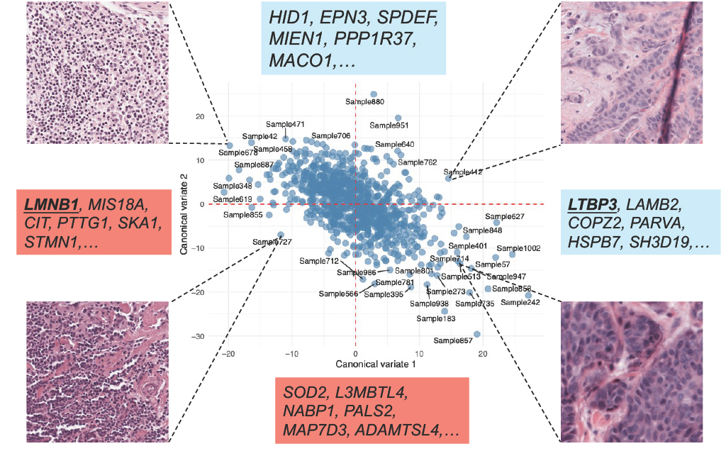
Integrating breast cancer tissue imaging and gene expression towards earlier disease diagnosis and individual treatments

A paradox sits at the heart of modern biology. A single tissue sample can now yield measurements on tens of thousands of genes, proteins, metabolites and microbes. And yet, the more we measure, the harder it becomes to draw meaning from the data. Conventional statistics were never designed for datasets where biological variables outnumber observations by orders of magnitude, and where the signal of interest sits inside vast and correlated noise. The bottleneck in modern life science is no longer measurement but interpretation.

Modern biological science is defined by data, but much of that data is too complex to analyse using conventional statistical tools.

Advances in genomics, proteomics, metabolomics, microbiome analysis and single-cell technologies allow researchers to measure tens of thousands of biological variables from a single sample.

While this has transformed what can be observed, it has also created a major analytical bottleneck.

Mapping global picoplankton biogeography

Traditional statistical methods struggle with these ‘high-dimension, low-sample-size’ datasets, where the number of variables (omics features) far exceeds the number of observations and where meaningful biological signals are embedded within large, highly correlated and noisy measurements.

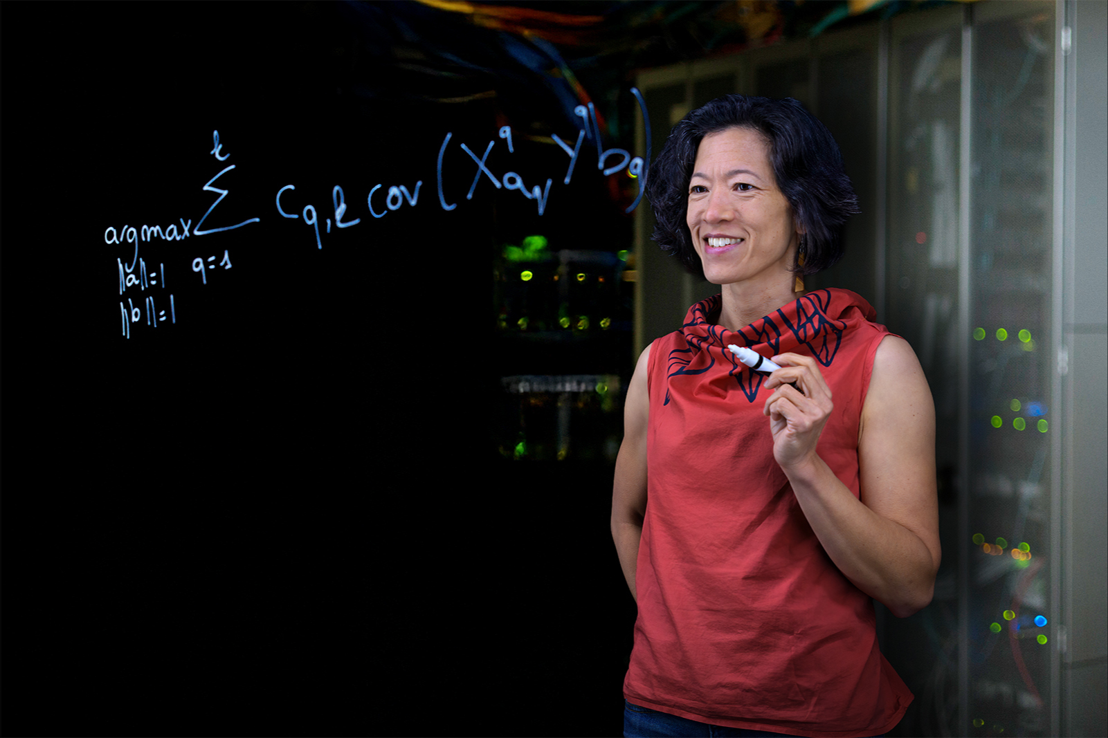
Prof Kim-Anh Lê Cao has spent more than a decade developing mixOmics to address this problem. The open-source suite brings together genes, proteins, metabolites, microbes and other ‘omics’ measurements within a single analytical framework, so that researchers can analyse them jointly rather than one layer at a time.

‘mixOmics methods give a holistic view of biological systems by integrating several layers of molecular information simultaneously, and identifying key molecular drivers in these complex systems,’ says Prof Lê Cao.

The impact of this capability is demonstrated through its application to real-world challenges across all life science.

  • In Australia’s dairy industry, mixOmics has been used to analyse complex milk metabolite profiles and to develop predictive models of cow fertility, underpinning more targeted breeding strategies, improved farm productivity and sustainability.
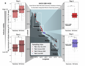
  • In environmental science, the Australian Institute of Marine Science applied mixOmics to integrate data from multiple Great Barrier Reef monitoring campaigns, enabling identification of microbial functional signatures that reliably predict water chemistry, providing a more sensitive framework for assessing reef health.
  • In human health, mixOmics supported integration of microbiome and metabolomic data to distinguish patients with chronic obstructive pulmonary disease, and to investigate disease susceptibility in a range of clinical contexts.
image of seawater analysis
mixOmics analysis accommodating seawater variation across different seasons

Across all these domains, the common outcome is not simply improved statistical performance, but the ability to turn previously intractable datasets into actionable biological insight.

MixOmics is currently being used by a large international research community of 50,000 users a year and its 13 methods developed by Prof Lê Cao and her team are widely cited and embedded in both academic and industrial research pipelines (9,000+ citations and 160+ patents using mixOmics).

Within QUBIC, mixOmics represents an enabling platform rather than a quantum technology in itself.

As quantum imaging and sensing systems begin to generate new forms of high-dimensional biological data, the analytical challenges they create will be similar in scale and complexity to those already addressed by mixOmics.

The established capability of the mixOmics platform positions QUBIC to interpret, integrate and translate quantum-derived biological information, supporting evidence-based decision-making and real-world impact as quantum-biotechnology matures.

The second generation of mixOmics, mixOmics PRO, has been registered as a company to further accelerate discoveries in omics life sciences.

QUBIC technologies are expected to generate complex data at the molecule, cell and tissue level with unprecedented time resolution. Methods such as those developed in mixOmics will extract insightful information from these different but complementary techniques from quantum sensors to sequencing experiments.

Feature image: Professor Kim-Anh Lê Cao. Credit: Mike Rennie Creative

Seeing and Controlling the Molecular Engines of Life

Imagine a future where medical treatments are more responsive, biological systems are easier to control, and disease can be detected earlier and more precisely. Reaching this future depends on understanding how life organises itself at the most fundamental, molecular level and how those processes might be guided or redesigned.

Many of the processes that sustain life occur at time and length scales far beyond what we can see. At the molecular scale, living systems organise themselves dynamically, forming temporary structures that control how cells function, adapt and respond to their environment. Understanding this hidden layer of organisation is essential for developing more effective therapies, diagnostics and biotechnologies.

Biomolecular condensates are emerging as a unifying framework for understanding and eventually shaping this molecular organisation.

Biomolecular condensates are dynamic, membrane‑less compartments that form when proteins and nucleic acids self‑assemble inside cells. Rather than being enclosed by physical boundaries, these structures arise through collective molecular interactions, allowing cells to concentrate and regulate biological activity with
remarkable flexibility.

Biomolecular condensates play a central role in organising life at the molecular level. They help regulate gene expression, coordinate biochemical reactions and
enable cells to respond rapidly to change. The same properties that make biomolecular condensates powerful biological tools also place them beyond the reach of many existing techniques.

Condensates are small, highly dynamic and governed by subtle molecular forces. Small changes in their composition or environment can significantly alter
their behaviour. In healthy systems, this adaptability is essential. In disease, however, condensates can become disrupted, contributing to conditions such as
neurodegeneration and cancer.

Understanding how condensates form and function, and how they might be controlled, requires new ways to measure molecular interactions with exceptional sensitivity.

Where quantum biotechnology enters the picture

Many of the key processes within biomolecular condensates occur at the nanoscale, where classical measurement tools struggle to capture weak and transient interactions. This is precisely the regime where quantum technologies offer new opportunities.

Ultra‑sensitive quantum sensors, advanced spectroscopic techniques and quantum‑informed simulations provide new ways to probe molecular organisation and dynamics. When combined with experimental platforms in molecular and cellular biology, these tools are allowing QUBIC researchers to characterise condensates with unprecedented precision.

QUBIC provides the environment where these capabilities come together, linking quantum science with biological experimentation and theory.

From insight to application

By learning how to control the formation and properties of biomolecular condensates, researchers could design programmable biomaterials with applications across
health and biotechnology, including:

  • Smarter drug delivery systems that respond dynamically to their environment
  • Synthetic bioreactors that organise complex reactions without rigid boundaries
  • New diagnostic platforms that exploit condensate sensitivity to molecular change

These possibilities show how quantum biotechnology extends beyond measurement, opening pathways to designing and engineering living matter itself.

A unique capability at the molecular frontier

Biomolecular condensates sit squarely within QUBIC’s mission to apply quantum technologies where biological complexity is greatest and new tools are most needed. By uniting researchers across institutions and research themes, the centre connects fundamental molecular insight directly to biological relevance.

This work positions QUBIC to drive future advances in healthcare, diagnostics and biotechnology by revealing how life organises itself at the molecular scale and turning that understanding into capability.

A centre‑wide effort across themes

In 2025, researchers from three QUBIC nodes (University of Wollongong, The University of Queensland, and University of Technology Sydney) published a major review in Advanced Materials: Biomolecular Condensates as Emerging Biomaterials: Functional Mechanisms and Advances in Computational and Experimental Approaches. Spanning the Molecules, Cells and Brain themes, the review integrates expertise in molecular physics, chemistry, biology and computation to examine biomolecular condensates from multiple perspectives. It brings together advances in experimental techniques and computational modelling to reveal the physical principles that govern condensate behaviour, and to explore how these systems could be developed as a new class of functional biomaterials.

This is precisely the kind of problem QUBIC exists to solve, because progress depends on integrating physics, chemistry, biology and computation in ways
that individual disciplines, projects or institutions cannot achieve alone.

Read more about QUBIC’s Molecules theme

An extract from the 2025 QUBIC Annual Report. Read the full report here.

Photonic molecular fingerprinting for fairer sports

Quantum assays for anti-doping control

The challenge

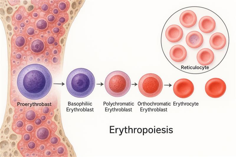
Erythropoietin (EPO) is a natural hormone that stimulates red-blood-cell production. Synthetic EPO closely mimics the natural form, making it hard to detect quickly and at low concentrations when misused for illegal doping. Current, lab-bound methods are slow and complex, creating a need for faster, more sensitive, field-ready tests.

The solution

A quantum photonic lab-on-a-chip that traps and analyses single EPO molecules, measuring mass, electrical charge and a vibrational fingerprint to tell natural from synthetic EPO. Combining these readouts aims to improve speed and sensitivity, potentially suitable for real-world, trackside testing.

The research

A QUBIC research team at the University of Queensland is developing and validating an integrated chip that optically traps proteins to provide label-free molecular fingerprinting of EPO at very low concentrations. “We start with known samples, set calibration, confirm specificity and detection limits, build and trial a prototype chip for anti-doping workflows,” says researcher Dr Igor Marinkovic.

Impact

“Direct molecular fingerprinting could redefine how EPO doping is detected,” says Dr Pavlina Naydenova. “Quantum photonic chips promise faster, more sensitive and selective testing, giving sporting bodies and clinicians a powerful new tool to safeguard athlete health and ensure competition integrity.”

Research team
  • Dr Igor Marinkovic
  • Dr Pavlina Naydenova
  • Dr Nicolas Mauranyapin
  • Prof Warwick Bowen
  • Kyle Clunies-Ross

Funded by the Queensland Government’s Quantum 2032 Challenge

Applying Quantum to the Brain Frontier

QUBIC CI Professor Marta Garrido brings expertise and infrastructure to enable next-generation quantum-enabled brain measurement.

The appointment of Professor Marta Garrido to QUBIC in 2025 marks a significant step in expanding the Centre’s multidisciplinary capability and strengthening its leadership in quantum-enabled biotechnology.

Her expertise in neuroscience and computational modelling brings new capability at the intersection of physics, engineering and biological research, supporting QUBIC’s future work in quantum-enabled human brain imaging.

Professor Garrido’s research combines brain imaging techniques and computational modelling to understand how the brain learns from experience and makes decisions in both typical individuals and people with psychiatric disorders. Her work uses methods including magnetoencephalography (MEG), electroencephalography (EEG), and magnetic resonance imaging (MRI) to study brain activity and neural circuitry. This research focuses on understanding the biological basis of brain function and psychiatric conditions, including disrupted predictive processes and brain circuitry.

Professor Garrido significantly expands QUBIC’s capability in magnetoencephalography.

She brings more than twenty years of experience in MEG data acquisition, analysis and brain connectivity modelling. In 2024, she established the first purpose-built whole- head, room-temperature MEG facility in the southern hemisphere at the University of Melbourne through an ARC LIEF grant. The facility uses wearable optically pumped magnetometers (OPMs) – 50 highly sensitive quantum magnetic field detectors – to measure extremely weak magnetic signals generated by brain activity. These sensors can be positioned flexibly on the head and allow recordings during more ‘natural’ experimental conditions where people can freely move.

This facility provides infrastructure to support QUBIC’s research in quantum-enabled neural imaging. In 2025, the University of Queensland also committed $1.6M to establish an R&D facility for quantum MEG. These two facilities combined means QUBIC has the southern hemisphere’s only room-temperature MEG facilities. Professor Garrido’s work contributes to informing the development of quantum sensors for MEG, benchmarking emerging sensing technologies including rubidium, diamond and optomechanical systems, and validating new approaches to non-invasive brain measurement. Her expertise in MEG data acquisition and analysis supports the development and testing of sensing technologies under development within QUBIC and contributes to research on brain connectivity and neural activity.

Her appointment also supports collaboration across QUBIC nodes. Project funding associated with this work enables the appointment of a postdoctoral research fellow in whole-brain MEG and will support MEG research at the University of Melbourne, University of Wollongong and The University of Queensland. Professor Garrido also brings experience in mentoring researchers, supporting training and development programs, established industry connections, and from 2026 will lead the QUBIC’s Outreach & Engagement portfolio.

Professor Garrido’s appointment strengthens QUBIC’s capacity to integrate quantum sensing technologies with biological and clinical research, expanding the Centre’s multidisciplinary capability and supporting collaboration across its research nodes. By contributing new infrastructure, technical expertise and research networks, her appointment enhances QUBIC’s ability to develop and apply quantum technologies for brain measurement and builds the foundation for future research and partnerships.


An extract from the 2025 QUBIC Annual Report. Read the full report here.

Reshaping How Scientists Explore the Physical Behaviour of Living Systems

Optical trapping and manipulation, which is the use of precisely shaped laser beams to hold, rotate, and measure microscopic structures, is reshaping how scientists explore the physical behaviour of living systems. These light‑driven tools allow researchers to quantify forces, observe rapid motions, and detect subtle mechanical changes inside cells at extremely small scales. Many of these measurements were previously out of reach, yet they are crucial for understanding how cells function, adapt and respond to stress or disease triggers. 

At The University of Queensland, the Rubinsztein‑Dunlop lab is at the forefront of this work. Dr Mark Watson is one of the researchers expanding what optical tweezers can do, having recently completed his PhD, Mark is continuing to advance the methods. His specialty lies in rotational and ballistic optical tweezers, which are platforms that can track the rotation of microscopic probes at exceptionally high speeds. These techniques allow scientists to capture dynamic processes in cell‑like environments on millisecond timescales, revealing biological activity that happens too quickly for many conventional tools to detect.

A major focus of Mark’s research is improving how we measure the physical properties of tiny biological environments. Using rotational optical tweezers he has developed targeted approaches to measure the fluid properties within cells by using light to control and twist a microscopic spherical probe. These measurements reveal physical signatures that provide clues about how cells move, change shape and perform their functions. Mark’s published studies show these tools in action in living cells and soft biological materials, highlighting their potential to uncover early signs of changes linked to health or disease.

Mechanical properties inside cells, such as viscosity, stiffness and force transmission, are central to processes including cell division, development, immune responses and disease progression. Tools that can measure these properties directly and in real time give scientists a window into how life operates at the smallest scales. Mark’s work enables exactly this – fast, sensitive and minimally disruptive measurements that can capture the physical “early warning signs” of how a cell is changing. These capabilities are essential for studying disease mechanisms, testing drugs and uncovering how subtle physical shifts influence biological behaviour.

As QUBIC develops quantum‑enhanced sensing and imaging technologies, high‑precision optical trapping systems will form a vital bridge between classical photonics and emerging quantum tools. Mark’s improvements to stability, bandwidth and calibration help ensure these platforms are ready for integration with quantum light sources and quantum‑enabled readouts, future techniques that promise to reveal biological processes with unprecedented sensitivity.

Continuing as a postdoctoral researcher in the Rubinsztein‑Dunlop lab, Mark is now extending his work into new regimes where optical manipulation is combined with faster detection, new forms of structured light and emerging approaches from quantum photonics. The application of his research is guided by close collaboration with cell biologists across QUBIC to determine which biological problems to tackle and what further developments are needed. These developments will contribute directly to QUBIC’s mission to create the next generation of precision tools for understanding life at its most fundamental scales.

Quantum Breakthrough Could Transform Heart Disease Diagnosis in Australia

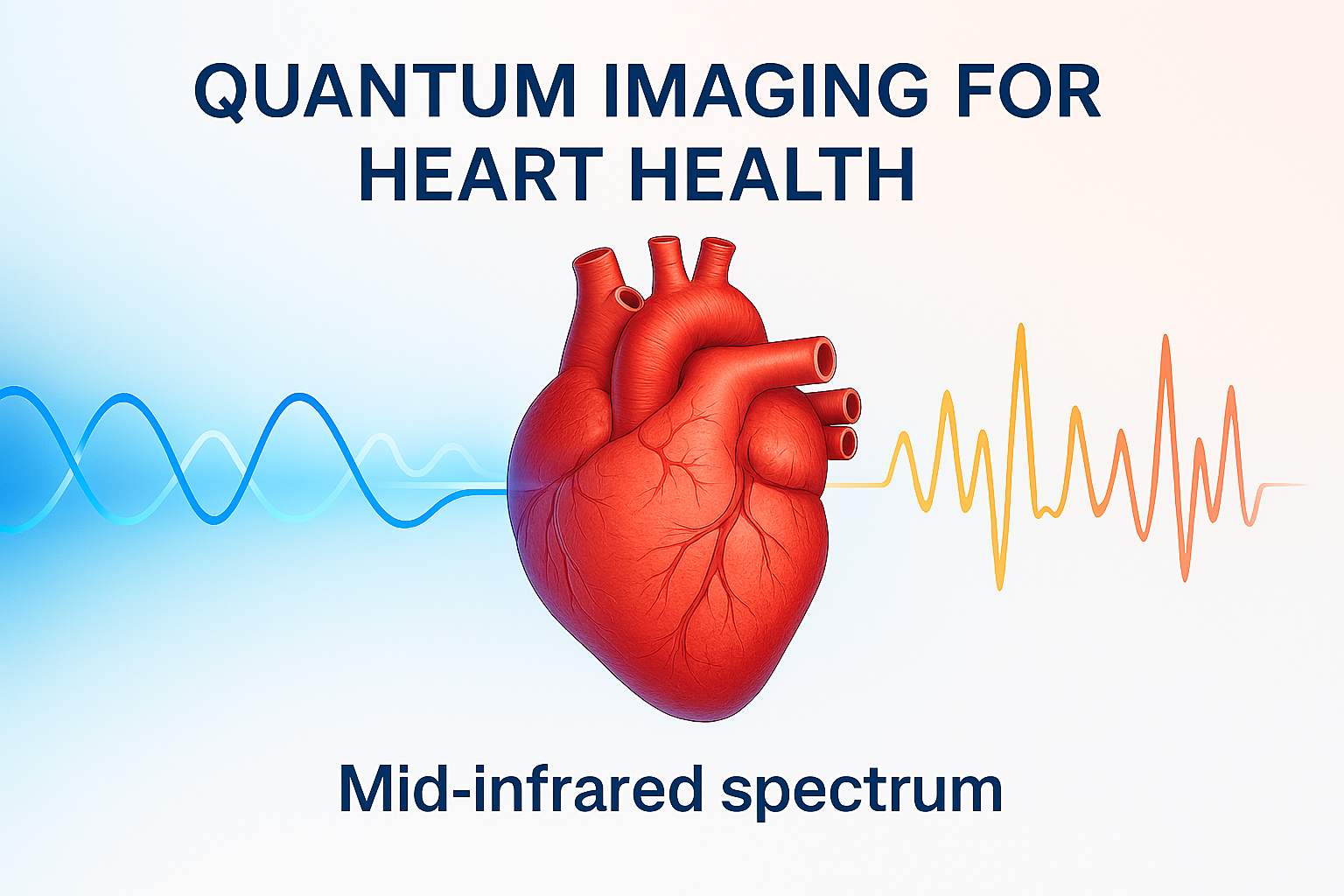
Heart disease remains the leading cause of death worldwide, and early, accurate diagnosis is critical to saving lives. Researchers at the ARC Centre of Excellence in Quantum Biotechnology (QUBIC) have taken a major step toward that goal by developing a quantum mid-infrared imaging approach that shows promise for distinguishing healthy from diseased heart tissue at the molecular level. This achievement, delivered through the Australian Government’s $36 million Critical Technology Challenge Program (CTCP), moves the technology closer to real-world use and positions Australia at the forefront of quantum-enabled health innovation.

The project demonstrates how quantum technologies can provide precise, non-invasive insights into heart health, paving the way for faster and more accurate diagnosis. It also marks progress toward prototype development, advancing beyond laboratory validation.

“By showing that quantum mid-infrared imaging can identify disease signatures in heart tissue, we’ve opened a pathway to practical diagnostic tools that could transform cardiovascular care,” says Professor Irina Kabakova, project lead at the University of Technology Sydney.

How the Technology Works
Infrared light can reveal the unique “fingerprints” of molecules because proteins, lipids, and other biomolecules absorb light at specific wavelengths. The mid-infrared range is particularly powerful for detecting these vibrational signatures, which change when tissue becomes diseased. QUBIC’s approach uses quantum-generated entangled photons to probe samples with high sensitivity and without dyes or labels. This enables label-free imaging and spectroscopy that can detect subtle molecular changes in tissue, such as those linked to heart disease, potentially allowing earlier and more accurate diagnosis.

The work is led by a cross-disciplinary team at UTS, including Professor Irina Kabakova, an expert in optical biomedical systems; Professor Alexander Solntsev, a leader in quantum optics and photonics; A/Prof Lana McClements, an expert in cardiovascular health, and Dr Isa Ahmadalidokht, who specialises in quantum spectroscopy and microscopy for diagnostics. Their combined expertise is enabling the transition from fundamental research toward applied health technology.

About the Critical Technology Challenge Program
The Critical Technology Challenge Program is a $36 million initiative under Australia’s National Quantum Strategy, designed to accelerate commercialisation of quantum technologies by moving them from early-stage readiness toward deployment and adoption. Round 1 Challenges included improving medical imaging and sensors for disease diagnosis, aligning directly with QUBIC’s heart disease spectroscopy project.

Bringing quantum to life
Quantum technologies are unlocking new frontiers in drug discovery, biomedical imaging, neuroscience and clean energy. Global investment in quantum technologies has already exceeded $55 billion, with the market projected to reach $106 billion by 2040. Life sciences are emerging as one of the most promising application areas, with quantum computing alone estimated to create $200–$500 billion in value by 2035, particularly through breakthroughs in drug discovery, diagnostics, and molecular simulation.

About QUBIC
The ARC Centre of Excellence in Quantum Biotechnology (QUBIC) is the world’s first national centre at the intersection of quantum science and biotechnology. QUBIC is developing next-generation quantum tools – including brain imagers and single-protein sensors – to tackle major challenges in health, biosecurity, energy, and agriculture. QUBIC’s research institutions include the University of Technology Sydney, University of Wollongong, The University of Queensland, the University of Melbourne, and Flinders University, and partners with leading industry, government, and international institutions.

Engineering Surfaces to Unlock Reliable Quantum Sensing

Quantum sensing promises measurement capabilities that far exceed classical technologies. Using quantum properties of matter, quantum sensors can detect extremely small magnetic, electrical and thermal signals, creating new opportunities across materials science, energy systems, health, advanced manufacturing, and the study of molecular and non‑equilibrium biological systems.

Quantum sensors are so sensitive that interference from their own surfaces overwhelms the signals they are designed to measure. Until this problem is solved, quantum sensing remains fragile, difficult to scale, and largely confined to controlled laboratory experiments.

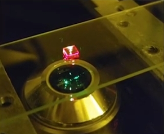
In work published in ACS Nano, QUBIC Chief Investigator Associate Professor David Simpson and collaborators directly addressed this challenge by engineering the surface of fluorescent nanodiamonds, which are a leading solid‑state quantum sensing platform.

Nanodiamonds containing nitrogen‑vacancy (NV) centres can operate at room temperature and offer nanometre‑scale spatial resolution. However, when these quantum defects are positioned close to the nanodiamond surface – a requirement for high‑resolution sensing – surface‑induced noise rapidly degrades performance. This surface noise has been a persistent barrier to practical quantum sensing.

By deliberately modifying nanodiamond surface chemistry and applying ultra‑thin, uniform silica coatings, the researchers suppressed surface‑generated noise and extended spin relaxation times into the millisecond regime, significantly improving the stability and performance of nanodiamond quantum sensors.

Crucially, this work demonstrates that stabilising quantum sensor performance can be achieved through materials engineering, without reliance on complex quantum control techniques. By clearly linking surface chemistry to quantum behaviour, Associate Professor Simpson and his team transformed surface modification from trial and error into a method that can be deliberately designed and optimised.

This capability is essential for quantum sensing in complex, dynamic environments that demand extreme spatial resolution, such as observing how new materials behave at the nanoscale, monitoring chemical reactions as they happen, improving energy technologies like batteries, and studying molecular‑scale processes as they unfold.

For QUBIC, this research strengthens Centre objectives by delivering a robust, scalable quantum sensing platform that underpins translation across biotechnology, including agriculture, biosecurity, clean energy, and health.

Read the paper here: Functionalized Fluorescent Nanodiamonds with Millisecond Spin Relaxation Times

Heat-Activated Imaging: New NIR-II Material Glows for a Longer Lifetime

Seeing deep into the brain without harming delicate tissue is one of the biggest challenges in medical imaging. Researchers from QUBIC at the University of Technology Sydney have developed a new material that could help, one that glows longer and more stably as temperatures rise.

Published in Nano Letters, the material emits long-lasting near-infrared (NIR-II) light, which is ideal for deep-tissue imaging. Unlike traditional materials that fade when they heat up, this one becomes more luminous, making it easier to see what’s happening inside the body, potentially useful during surgery or in areas where temperature changes are common.

This research supports QUBIC’s mission to develop quantum-enabled technologies that reveal the inner workings of living systems. The material leverages the quantum properties of lanthanide ions, pairing special energy levels of different ions to enable more efficient energy transfer at higher temperatures. This design turns thermal fading, a long-standing problem, into an advantage, allowing for clearer, more stable imaging when it’s needed most.

By advancing the fundamental understanding of energy transfer in lanthanide systems, this work contributes to the development of next-generation imaging materials that could support neurological research, where non-invasive, high-resolution access to brain structures is critically needed.

Published paper: Thermally Prolonged NIR-II Luminescence Lifetimes by Cross-Relaxation (2024)

Cover of the 2024 Annual ReportThis impact story is an extract from QUBIC’s 2024 Annual Report: read more.